
In-House Lab & Diagnostics
Diagnostic Imaging Services
At Arkansas Veterinary Emergency & Specialists, we provide diagnostic imaging including x-ray, ultrasound, and CT scan for pets in Little Rock. These technologies produce highly detailed images of the internal structures of your pet's body to help provide accurate diagnosis.
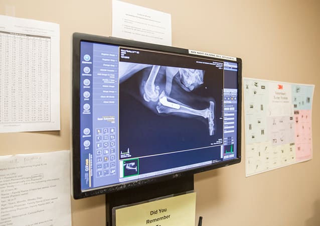
Radiography (Digital X-rays)
A radiograph, or x-ray, is a type of photograph that can look inside the body and reveal information that may not be discernible from the outside.
Radiography is painless, safe, and completely non-invasive, and uses only very low doses of radiation. Because the level of radiation exposure needed to perform radiography is very low, even pregnant females and very young pets can undergo radiography.
Radiographs can be used to evaluate bones and organs and diagnose conditions including bladder stones, broken bones, chronic arthritis, spinal cord diseases and some tumors.
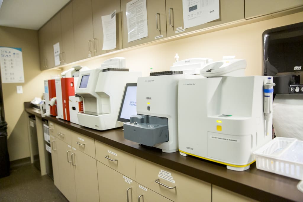
In-House Lab & Pharmacy
Our in-house laboratory allow us to perform tests and get results quickly, so that we can diagnose your pet's symptoms and start treatment as soon as possible. Our pharmacy is stocked with a range of medications and prescription diets, giving us quick access to any medications your pets may need while in our care.


Ultrasound
Ultrasound is a non-invasive technology that uses sound waves transmitted into the body to create an image that allows your vet to look at the architecture of an organ.
With ultrasound, fluid can be distinguished from soft tissue masses or foreign bodies, which can be difficult to do with a digital x-ray. This makes the ultrasound useful in the diagnosis patients for hemoabdomen and pericardial effusion (blood in the abdomen and around the heart).
When a pet has a tumor or ingests something it shouldn’t have, an ultrasound can also be helpful to locate the objects and characterize them.
Computed Tomography (CT scan)
A computed tomography (CT) scanner is an advanced, non-invasive and painless diagnostic imaging tool that allows our veterinarians and specialists to view the internal anatomy of a patient by producing highly detailed cross-sectional images.
Our veterinarians frequently use CT scans for planning surgery and for diagnosing specific disease processes that affect certain regions in the body, such as the spine, nasal cavity, middle/inner ear, abdomen, thorax, and joints.
From Our Clients
-
"This place was awesome. The staff was wonderful. They explained everything to us, took amazing care of our fur baby and kept us updated. They even gave me an update (I even spoke directly to the vet) when I called at 1:30 in the morning! I would recommend this place to anyone."